
Target the right cells. Deliver the right therapy. Change the outcome.
Introducing Jiksak’s Neuron Drug Delivery System (Neuron DDS).
This innovative drug delivery technology enables precision, high concentration delivery of drugs to target neurons. Thus efficiently delivering them to the central nervous system within the brain and spinal cord.

The key challenge in neurodegenerative diseases is not discovering new drugs, but delivering them to the right neurons that degenerate. Random uptake by healthy cells can cause harmful off-target effects, while too little drug reaches diseased neurons to make a difference—leading many CNS drug candidates to drop-out at this critical step.
Our drug delivery platform exploits a unique physiological gateway to deliver therapies directly to neurons, bypassing the central nervous system barriers that block effective drug delivery. The outcomes are improved safety, enhanced efficacy, and a real opportunity to transform scientific innovation into real patient care.
Motor neurons are among the body’s longest and most specialized cells, stretching from the spinal cord to the muscles. They constantly relay signals, maintaining a natural highway between the central and peripheral nervous systems. What if we could ride on this communication pathway to deliver drugs directly into the brain and spinal cord by entering through the muscle junction side located at the periphery? At Jiksak, we have turned this biology into a therapeutic opportunity.
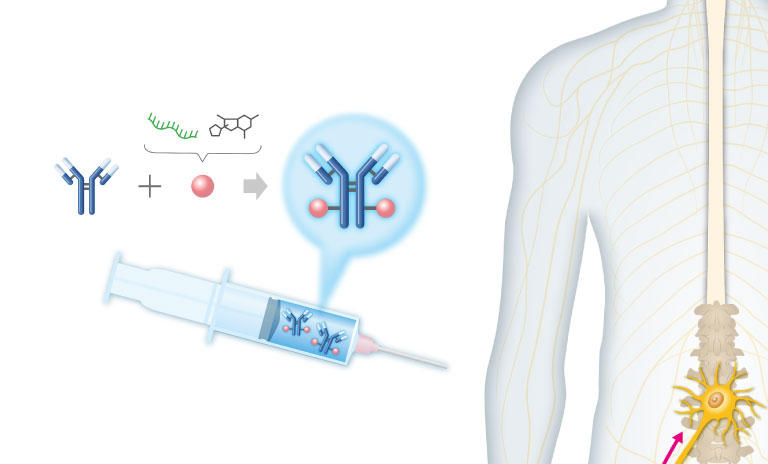
Step 1 Jiksak's Neuron DDS antibody seamlessly combines neuron targeting with therapeutic drugs, such as oligonucleotides and small molecules, for intravenous delivery.

Step 2 The antibody searches for motor nerve terminals by recognizing Synaptotagmin 2 (SYT2), a protein expressed on synaptic vesicles that store neurotransmitters. Transduction of brain signals to motor neuron (MN), releases neurotransmitters for muscle contraction. Upon the release of neurotransmitters, SYT2 is exposed to the junction space. This creates a fleeting and frequent window for antibody binding. Once bound, the antibody-drug complex is carried into the motor neuron through synaptic vesicle recycling; a natural process that neurons use to reset and reuse their signaling machinery.
In this way, our Neuron DDS platform delivers therapeutic molecules directly into motor neurons via their terminal ends.

Step 3 Once inside the nerve terminals, the DDS antibody transports the drug along the motor neuron’s internal transportation highways, the axons*, to reach the cell body deep in the spinal cord and brainstem. From there, it can distribute to other SYT2-positive neurons across the central nervous system (CNS)— expanding its therapeutic reach and unlocking the potential to treat widespread neural networks.
*Our company‘s name, Jiksak, means axons in Japanese embodies the assets and connections that define our company.
This unique mechanism turns a brief biological window into a powerful therapeutic gateway—enabling neuron-targeted delivery into the CNS without the need to physically cross its protective barriers.


